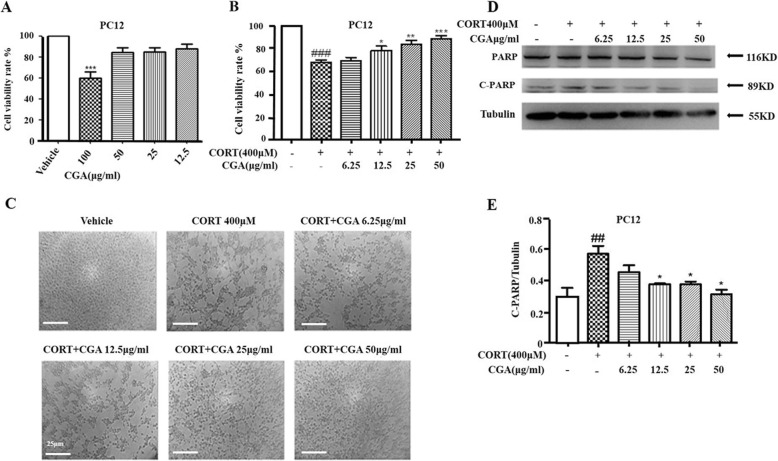
Fig. 2.
CGA protected against CORT-induced neurotoxicity in PC12 cells. a PC12 cells were cultured with various concentrations of CGA for 24 h, and cell viability was measured by MTT assay (n = 3). b Cell viability was determined by MTT assay (n = 3). c Changes in the morphology of PC12 cells were observed by optical microscope (Scale bar, 25 μm). d Changes in the expression of C-PARP were examined by Western blot. e Densitometric values of C-PARP were quantified using the AlphaEaseFC software. Data are presented as mean ± SD. # P < 0.05, ## P < 0.01, ### P < 0.001 compared with the vehicle group. * P < 0.05, ** P < 0.01, *** P < 0.001 compared with the CORT group

