
Fig. 2.
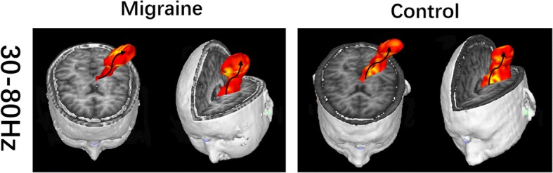
Real time source images showing spatial activities under somatosensory stimulation in gamma band (30–80 Hz) recorded from migraine patients and control. 3D images are displayed in axial, coronal and oblique sagittal positions. The black arrow indacates the source activity flow from the deep brain to the sensory cortex