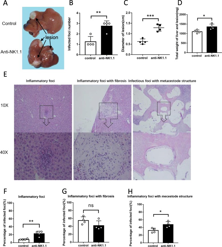
Fig. 3.
Hepatic histopathological alterations and granulomatous response in E. multilocularis infection while in vivo depletion of NK cells population. a The macroscopic views of the hepatic lesions infected mice. b The number of intrahepatic E. multilocularis infected lesions. c The sum of diameters for lesions in liver. d Total weight of the liver and infected hepatic lesions. e Histopathological alterations in liver of infected mice. H&E staining of liver sections. The original magnification was at 10×, and the below corresponding images were magnified at 40×, respectively. f, g and h Hepatic granulomatous response to E. multilocularis infection [12]. Data were shown as mean ± standard error (SEM, 4 mice per group), *p < 0.05. **p < 0.01, ***p < 0.001

