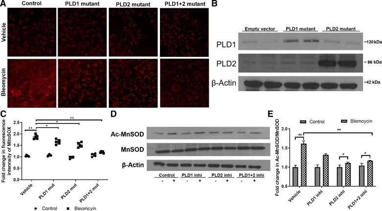
Fig. 6.
Inhibition (Inhi) of phospholipase D1 (PLD1) and PLD2 activity attenuates bleomycin-induced mitochondrial reactive oxygen species (ROS) generation and acetylation (Ac-) of manganese-superoxide dismutase (MnSOD). Bronchial airway epithelial (Beas2B) cells grown in 35-mm dishes (~60% confluence) were infected with adenoviral control or catalytically inactive PLD1 [50 MOI (multiplicity of infection)], PLD2 mutant (mut; 50 MOI), or a combination of PLD1 + PLD2 (50 MOI each) for 24 h before bleomycin challenge (10 mU/ml) for 1 h. Cells were then incubated with MitoSOX red reagent for 15 min and later subjected to 2 washes with phenol red-free medium. A: representative images of MitoSOX staining, a measure of mitochondrial superoxide (O2·−) generated. B: Western blot showing overexpression of PLD1 and PLD2 in Beas2B cells after infection with catalytically inactive mutants. C: quantification of fluorescence intensity of MitoSOX staining by ImageJ. D: Beas2B cells grown in 35-mm dishes (~90% confluence) were pretreated with 250 nM PLD1 inhibitor (VU0155609), 500 nM PLD2 inhibitor (VU0364739), or 250 nM VU0155609 + 500 nM VU0364739 for 3 h before bleomycin challenge (10 mU/ml) for 1 h. Cell lysates (30 µg protein) were subjected to Western blotting and stained for Ac-MnSOD and total MnSOD with antibodies. Shown is a representative blot from 3 independent experiments. E: quantification of Western blots (D) by densitometry/ImageJ analysis, and fold changes were normalized to total MnSOD. Values are means ± SD of 3 independent experiments. *P < 0.05, **P < 0.005.

