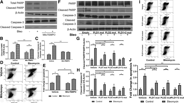
Fig. 8.
Inhibition of mitochondrial superoxide generation and phospholipase D (PLD) activity attenuates bleomycin (Bleo)-induced apoptosis in bronchial airway epithelial (Beas2B) cells. Beas2B cells grown in 35-mm dishes (~60% confluence) were infected with vector, PLD1 [50 MOI (multiplicity of infection)], PLD2 (50 MOI), or a combination of PLD1 + PLD2 mutants (mut; 50 MOI each) for 24 h. Cells were treated with MitoTempo (100 μM) for 1 h and challenged with bleomycin (10 mU/ml) for 24 h. Programmed cell death following bleomycin challenge was determined by cleavage of caspase-3 and polyADP ribose polymerase (PARP), indicators for apoptosis, and flow cytometry of propidium iodide (PI)/annexin V-positive cells. A: protein expression of caspase-3 and PARP in cell lysates (20 µg of protein) from cells with or without MitoTempo pretreatment. Shown is a representative blot from 3 independent experiments in triplicate. B and C: quantification of cleaved caspase-3 and PARP from A by densitometry and ImageJ analysis; data were normalized to β-actin levels. D and E: Beas2B cells grown in 35-mm dishes (~90% confluence) were exposed to bleomycin in the absence or presence of MitoTempo as in A, and apoptosis was quantified by flow cytometry analysis. Early apoptotic and late apoptotic cells were sorted out by labeling the cells with annexin V and PI and quantified. F–H: cell lysates from Beas2B cells infected with empty vector, PLD1, PLD2, and PLD1 + PLD2 mutant with and without bleomycin challenge as in A were analyzed by Western blotting for cleaved PARP and caspase-3 and total β-actin. Band intensities were quantified by densitometry and ImageJ and normalized to total actin. Shown is a representative blot from 3 independent experiments. I: Beas2B cells transfected with PLD1 or PLD2 mutants or PLD1 +PLD2 mutants as in F in the presence or absence of bleomycin (10 mU/ml) for 24 h were analyzed by flow cytometry for apoptosis. Shown is a representative dot plot of annexin V and PI staining. Late-apoptotic cells stained as annexin V+/PI+ cells seen on C++, and early-apoptotic cells were stained as annexin V+/PI− cells seen on C+/−. J: percentage of late-apoptotic cells were quantified from I. *P < 0.05, **P < 0.005.

