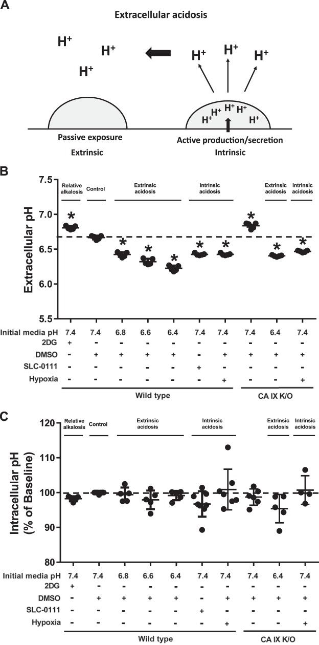
Fig. 1.
Twenty-four hours after extrinsic and intrinsic extracellular acidosis exposure, pulmonary microvascular endothelial cells (PMVECs) maintain intracellular pH. A: schematic representation of extrinsic and intrinsic extracellular acidosis: extrinsic acidosis is determined by passive extracellular proton exposure, and intrinsic acidosis is determined by increased cellular proton production and/or secretion. B and C: wild-type and carbonic anhydrase (CA) IX knockout (K/O) PMVECs were seeded at 5.0 × 10⁵ cells/well on 6-well plates (B) or 4.0 × 10⁴ cells/well on 96-well plates (C) on bicarbonate buffered media. Two days after cell seeding, media were changed to HEPES-buffered media with pH 7.4, 6.8, 6.6, and 6.4 and treated with 2-deoxy-d-glucose (2DG; 5 mM), DMSO (0.5%), and SLC-0111 (150 µM) in normoxia and hypoxia (1% oxygen). Extracellular acidosis is attenuated with 2DG treatment but enhanced with SLC-0111 or hypoxia (B). No significant change occurs in intracellular pH levels 24 h after extrinsic or intrinsic acidosis in PMVECs (C). Data represent means ± SD. Two-way ANOVA and Bonferroni post hoc tests were used to compare between different groups. At least five separate experiments were performed. *Significant difference (P < 0.05) from wild-type pH 7.4.

