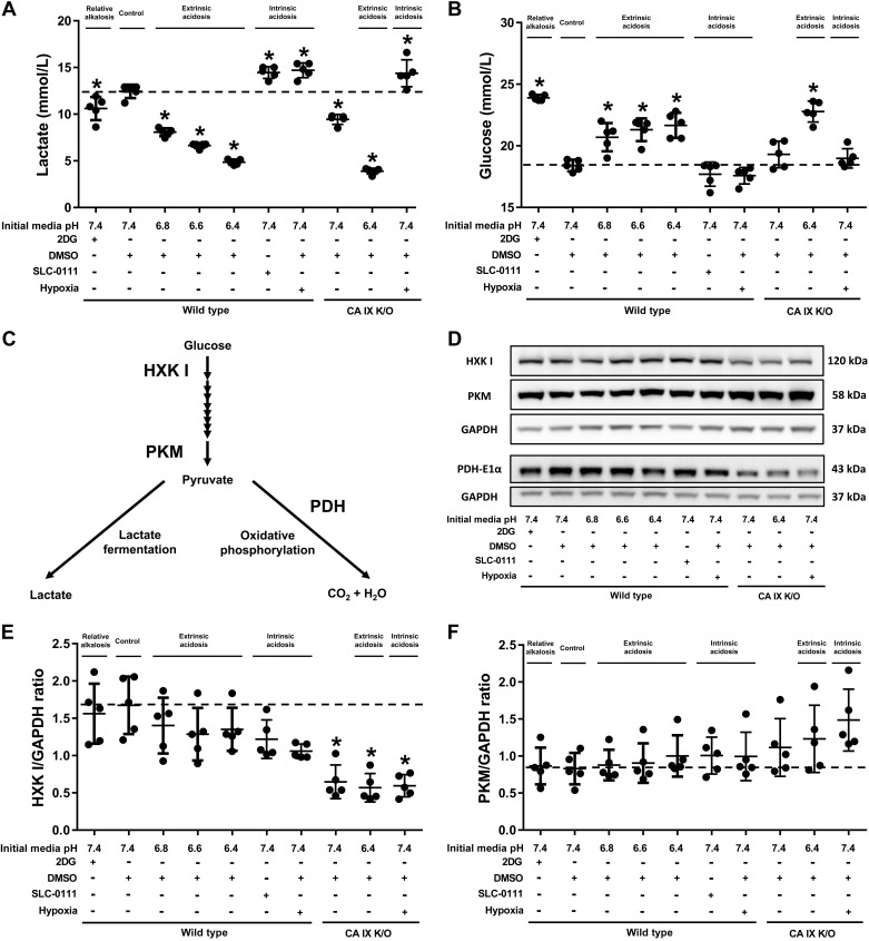
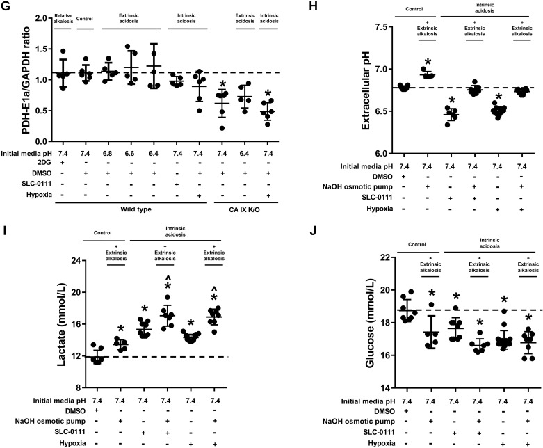
Fig. 3.
Extrinsic acidosis suppresses glycolysis. Wild-type and carbonic anhydrase (CA) IX knockout (K/O) pulmonary microvascular endothelial cells (PMVECs) were seeded at 5.0 × 10⁵ cells/well on 6-well plates on bicarbonate-buffered media. Two days after cell seeding, media were changed to HEPES-buffered media with pH 7.4, 6.8, 6.6, and 6.4 and treated with 2-deoxy-d-glucose (2DG; 5 mM), DMSO (0.5%), and SLC-0111 (150 µM) in normoxia and hypoxia (1% oxygen). Twenty-four hours later, media were collected for lactate and glucose measurements (A and B), and whole cell lysates were collected for Western blot analysis of main metabolic enzymes (C) pyruvate kinase M-type (PKM), hexokinase I (HXK I), and pyruvate dehydrogenase E1α subunit (PDH-E1α) (D–G). Extrinsic acidosis decreased lactate production and glucose consumption, while intrinsic acidosis increased lactate production (A and B). CA IX K/O PMVECs have decreased baseline lactate production and HXK I and PDH-E1α expression (A, B, E, F, and G). The same experimental setting was used to test the effect of extrinsic alkalosis on wild-type PMVECs in the presence of intrinsic acidosis, utilizing an osmotic pump to deliver NaOH (1 N NaOH 1 µl/h for 24 h). Media pH (H), lactate (I), and glucose (J) were measured, revealing glycolysis-enhancing effect of extrinsic alkalosis. Data represent means ± SD. Two-way ANOVA and Bonferroni post hoc tests were used to compare between different groups. At least five separate experiments were performed. *Significant difference (P < 0.05) from wild type pH 7.4.


