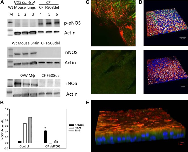
Fig. 2.
Endothelial nitric oxide synthase (eNOS) expression in human airway pseudostratified epithelium. A and B: ciliated air-liquid interface (ALI) culture membranes from an F508Del cystic fibrosis (CF) transmembrane regulator (CFTR) homozygous patient (2 filters/lane) and wild-type (WT) mouse lungs’ tissue were immunoblotted for phospho-eNOS S1177 (top). In a separate experiment, membranes from an F508Del CFTR homozygous patient and WT mouse brain’ tissue were immunoblotted for neural nitric oxide synthase (nNOS; middle) and inducible nitric oxide synthase (iNOS; bottom). RAW cells macrophages served as a control for iNOS. Each membrane was stripped and re-probed for β actin (ALI 1 and 2). B: phospho-eNOS immunostaining was greater than for iNOS and nNOS in CF F508Del (*P < 0.001). C: eNOS staining from an ex vivo epithelial prep from A underwent immunofluorescent staining. C, top: eNOS (red) and dynein (green) are apical. C, bottom: negative control image shows no autofluorescence (ALI 7). D: ciliated ALI cultures from a non-CF subject (ALI 5) were immunostained for eNOS (top, green) or isotype control (bottom). Background was stained for filamentous actin (red), ciliary tubulin (white) and nuclei (blue). Sparse apical eNOS is observed. E: ciliated ALI cultures from a F508Del homozygous patient (ALI 2) were immunostained for eNOS (red), which is near apical and at times colocalized with dynein (green). Note sparse apical staining and absence of colocalization with the nucleus (DAPI; blue) or basal cells.

