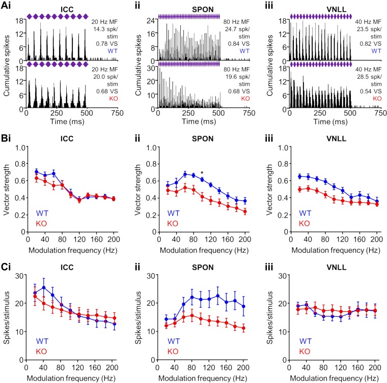
Fig. 9.
Temporal acuity to envelope modulation responses of neurons in α7-knockout (KO) mice. A: representative wild-type (WT) and KO responses in the inferior colliculus (ICC; i), superior paraolivary nucleus (SPON; ii), and ventral nucleus of the lateral lemniscus (VNLL; iii) to sinusoidal amplitude modulation (SAM) of the sound envelope. Purple bars represent 500-ms characteristic frequency (CF) tones presented at 20 dB above threshold with 100% modulation depth at various modulation frequencies (MF). Response fidelity to individual cycles of modulation was determined by the vector strength (VS; see materials and methods). B: lower vector strengths for KO neurons in the ICC (i), SPON (ii), and VNLL (iii), particularly at low modulation frequencies, were not significant in most cases (except SPON 100 Hz: P = 0.03, Kruskal-Wallis test/Dunn’s multiple comparisons test). C: no difference was found between spiking rates of WT and KO neurons in the ICC (i) and VNLL (ii). Spiking rates were consistently lower to most modulation frequencies for KO neurons in the SPON, but the differences did not reach statistical significance (P > 0.1 for each condition, Kruskal-Wallis test/Dunn’s multiple comparisons test). *P < 0.05.

