
Figure 2.
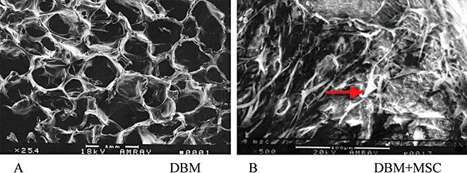
SEM photomicrograph of a cross‐section of DBM only (A) (original magnification ×25.4) shows the porous structure. After MSC were seeded on a DBM scaffold for further culture for five days in vitro, an SEM image of tissue‐engineered bone (B) (original magnification ×500) shows there are considerable numbers of cells (arrow) in the pores of the DBM scaffold.