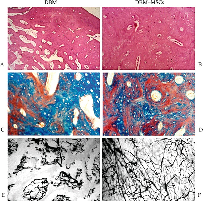
Figure 5.

Histological analysis of newly formed bone by HE staining (A, B), Masson staining (C, D), and Indian ink staining (E, F) at 12 weeks post surgery. The amount of newly formed bone in the DBM group (A) was less than in the DBM plus MSC group (B). However, the formation of fibrous tissue in the DBM group was more. An endochondral ossification pattern was observed in both DBM plus MSC and DBM groups, and cortical bone had formed around newly formed woven bone in the DBM plus MSC group. Compared with the DBM group, the more newly formed vessels were observed in the DBM plus MSC group by methods of Masson staining (D) and Indian ink staining (F).
