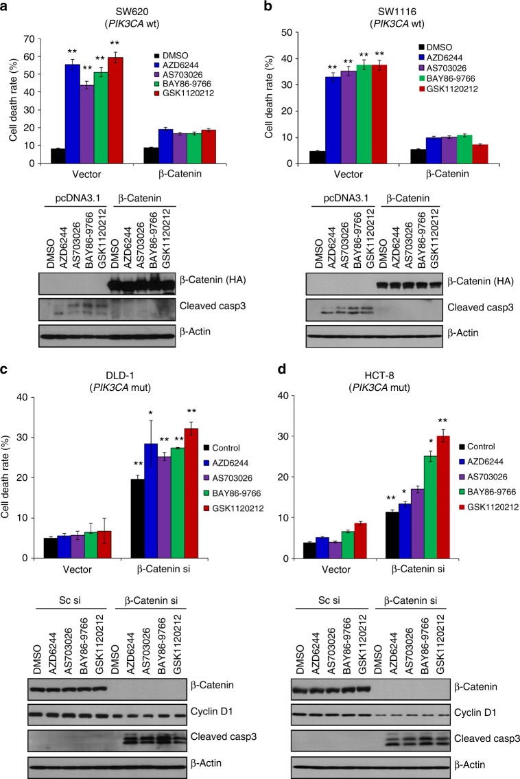
Fig. 2.
β-Catenin expression determines MEK inhibitor sensitivity. SW620 (a) and SW1116 cells (b) expressing wild-type PIK3CA were transfected with pcDNA3.1 plasmid containing empty vector or β-catenin for 24 h and then treated with various MEK inhibitors (1 µM) for another 24 h. a, b (Upper panel) Cell death was determined with the Trypan blue exclusion assay. The data represent the means ± SDs of at least three independent experiments. a, b (Lower panel) All resulting data were statistically analysed using a two-tailed Student’s t-test *P < 0.05, **P < 0.01. Cell lysates were used for western blot analysis using antibody targeting the HA tag or cleaved caspase-3. β-Actin was used as a loading control. DLD-1 (c) and HCT-8 PIK3CA mt cells (d) were transfected with scramble or β-catenin siRNA for 24 h and then treated with the indicated MEK inhibitors for another 24 h. c, d (Upper panel) Cell death was evaluated with the Trypan blue exclusion assay. The data represent the means ± SDs of at least three independent experiments. c, d (Lower panel). Western blot analysis was performed using antibodies targeting β-catenin, cyclin D1, cleaved caspase-3, and β-actin, which was used as a loading control. All resulting data were statistically analysed using a two-tailed Student’s t-test. *P < 0.05, **P < 0.01

