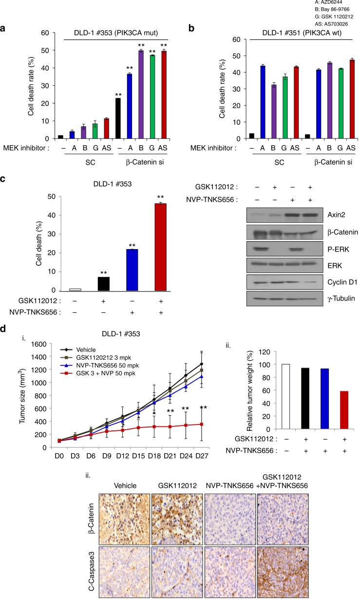
Fig. 4.
Inhibition of β-catenin in DLD-1–PIK3CA isogenic cells is determined by their sensitivity to MEK inhibitors. DLD-1–PIK3CA mt (cell line 353) (a) and DLD-1–PIK3CA wt (cell line 351) isogenic cells (b) were transfected with scramble siRNA or β-catenin siRNA for 48 h and then treated with the indicated 1 µM MEK inhibitors for another 24 h. MEK inhibitor resistance was overcome by β-catenin knockdown in PIK3CA mutant cells as shown by the Trypan blue exclusion assay. The data represent the means ± SDs of at least three independent experiments. All resulting data were statistically analysed using a two-tailed Student’s t-test. *P < 0.05, **P < 0.01 c Combinatorial effect of the MEK inhibitor GSK112012 and the β-catenin pharmacological inhibitor NVP-TNKS656 on DLD-1-PIK3CA mt (cell line 353) cells were evaluated with the Trypan blue exclusion assay. Cell lysates were used to analyse Axin2, β-catenin, p-ERK, ERK, and cyclin D1 expression by western blot analysis. γ-Tubulin was used as a loading control. All resulting data were statistically analysed using a two-tailed Student’s t-test. *P < 0.05, **P < 0.01. d Tumour from a nude mouse subcutaneously injected with DLD-1-PIK3CA mt (cell line 353) cells. (i) When the tumour volume reached 100 mm3, the tumours were treated daily with 3 mg/kg GSK112012, an MEK inhibitor, and/or 50 mg/kg NVP-TNKS656, a tankyrase inhibitor. Tumour size was measured every 3 days after drug treatment. The data represent the means ± SDs. Each group comprised 4–5 mice. All resulting data were statistically analysed using a two-tailed Student’s t-test *P < 0.05, **P < 0.01. (ii) Tumour weight was assessed after 27 days of drug treatment. (iii) β-Catenin and cleaved caspase-3 expression was confirmed by immunohistochemical analysis

