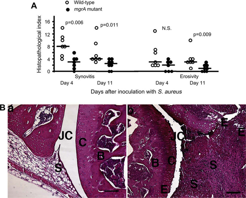
Fig. 4.
(A) Histologic evaluation of joints from all four limbs of NMRI mice, sacrificed 4 days (n = 7/group), and 11 days (n = 7 and 8 mice/group respectively) following i.v. inoculation with 3 × 10⁶ CFU of S. aureus Newman or its isogenic mgrA mutant. Comparisons were made using the Mann–Whitney U-test, NS, not significant. (B) (a) Micrograph showing an apparently intact knee joint from a NMRI mouse, 11 days after inoculation with 3 × 10⁶ CFU of the mgrA mutant. (b) Micrograph showing the knee joint from a mouse inoculated with the same dose of the wild-type strain Newman, displaying severe inflammation of synovial tissue and bone and cartilage erosion, JC, joint cavity; C, cartilage; B, bone; S, synovial tissue; E, erosion of bone and cartilage. Bar, 100 μm. Hematoxylin/eosin staining was used.

