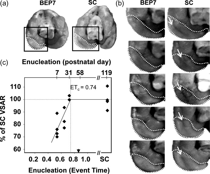
Figure 2.
Primary visual cortex surface area as a function of age of deprivation onset in bilaterally-enucleated ferrets. (a) The primary visual cortex resides on the medial occipital surface, indicated by dashed white boundaries in caudal/ventral views of the brain in a BEP7 and a sighted control (SC) ferret. Although the primary visual cortex in ferrets extends dorsally, the surface area reduction in brains of BEP7 ferrets is notable in the ventral cortex due to the absence of the occipital-temporal sulcus. (b) The surface area of the primary visual cortex (enclosed by dashed boundaries) of BEP7 ferrets is reduced compared with controls. The absence of the occipital-temporal sulcus in BEP7 animals (arrows in control brains), contributes to the difference in visual cortex surface area. (c) Quantification of VSAR by MRI revealed pronounced reductions in early enucleates (P7, ET = 0.54), with graded effects of enucleation on the primary visual to non-visual surface area ratio for later enucleations (P20, ET = 0.65 and P31, ET = 0.72). The Solid line indicates best fit of the piece-wise regression (Adjusted R2 = 0.64). Vertical dotted line indicates the estimated end of the effect of enucleation (ETc = 0.74). Arrowhead indicates V1 peak synaptic density. Abbreviations as in Fig. 1.

