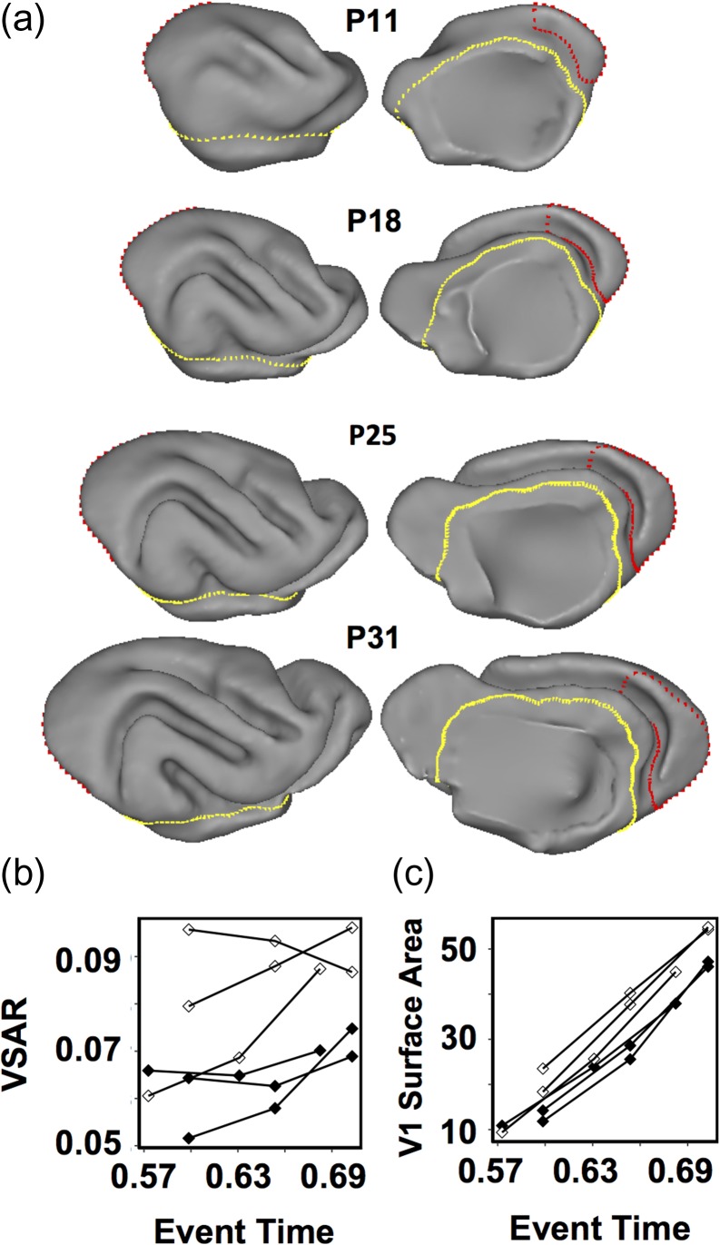
Figure 5.
Longitudinal MRI anatomical analysis in three sighted control ferrets and three ferrets enucleated at P7. The visual cortical surface area is reduced in the immature brain of P7 enucleated ferrets, compared with that in immature brains of control ferrets. (a) Lateral (left) and medial (right) views of the right hemisphere of a control ferret brain at ages P11, P18, P25, and P31. Gyri and sulci appear as they do in a mature brain, which enables delineation of striate visual cortex (red), as well as isocortex (yellow) boundaries following previously established conventions (Bock et al. 2012). (b) VSAR plots of the average of the left and right primary visual cortex surface area for three control (open symbols) and three BEP7 (black symbols) ferrets illustrating the reduction in surface area in the BEP7 ferrets induced by the enucleation. (c) Plots for the same animals, as in b, but showing V1 surface area (mm2) expansion over time.

