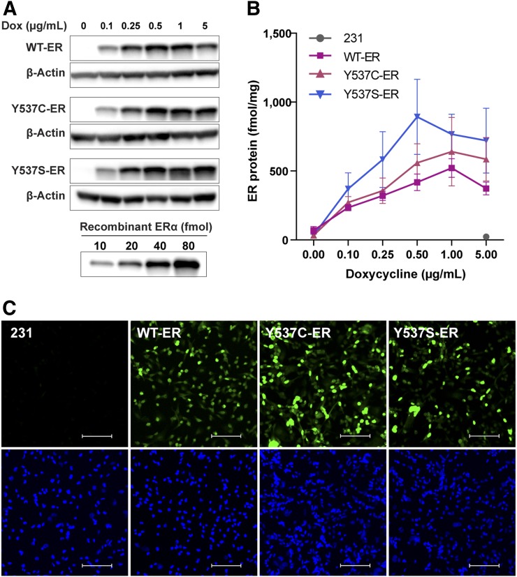
FIGURE 1.
ER protein expression and localization in generated breast cancer cell lines. (A) Representative Western blot of ER protein in WT-ER, Y537C-ER, and Y537S-ER cells treated with increasing doses of doxycycline for 24 h. (B) ER protein quantification (mean ± SEM) from 3 independent experiments. (C) Immunofluorescence for ER localization: Alexa fluor 488 staining for ER (top) and DAPI nuclear staining (bottom). Scale bar = 100 μm. Images are representatives of 3 individual experiments.

