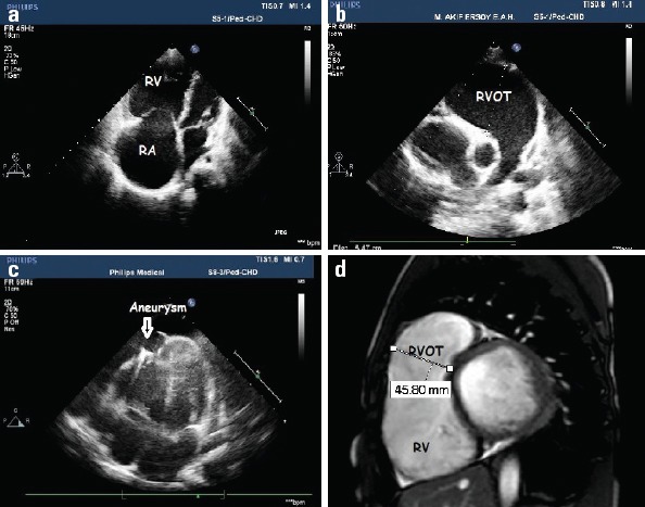
Figure 2.

Two-dimensional echocardiography and cardiac magnetic resonance images of patients with ARVD. (a) Two-dimensional echocardiographic image demonstrated dilatation of RV and RA. (b) Two-dimensional echocardiographic image demonstrated significant dilatation of RVOT. (c) Two-dimensional echocardiographic image demonstrated a localized right ventricular apical aneurysm in a patient with ARVD. (d) Cardiac MRI image demonstrated significant dilatation of RVOT and thinning of the walls in the RV myocardium in the sagittal plane
ARVD - arrhythmogenic right ventricular dysplasia, MRI - magnetic resonance imaging, RA - right atrium, RV - right ventricle, RVOT - right ventricle outflow tract
