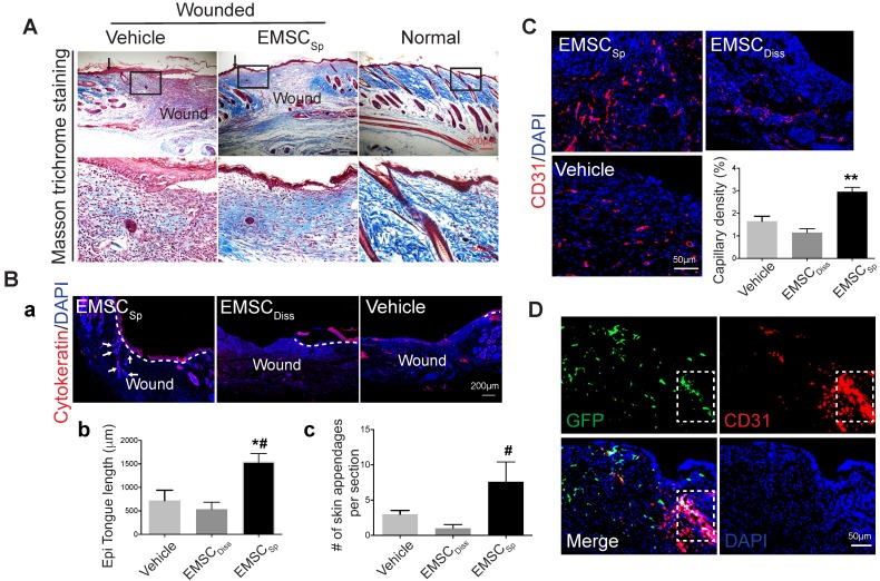
Figure 3.
Histological analyses of wounds with transplanted EMSCSp. (A) Masson trichrome staining of normal skin and wounds 14 days after treatment with EMSCSp or vehicle control. Boxed areas are amplified and shown on the bottom row. Arrows in the upper row indicate the junction between the wound and surrounding skin. (B) Re-epithelialization of wounds 14 days after treatment with EMSCSp, EMSCDiss or vehicle. Tissues isolated from the wounds were sectioned and immunostained (a) with anti-pancytokeratin antibody. White dashed lines indicate the detected epidermal keratinocytes, and white arrows indicate skin appendages. The epithelial tongue length (b) and the number of skin appendages per section (c) are shown in the bar charts. n = 5 biological repeats; *P < 0.05 for EMSCSp versus vehicle and #P < 0.05 for EMSCSp versus EMSCDiss per ANOVA followed by Tukey's test. (C) Detection of vascular endothelial cells in wounds 14 days after treatment as described above. Tissues isolated from the wounds were sectioned and immunostained with antibodies against CD31 as a marker for vascular endothelial cells. The results are quantified in a bar graph and displayed as capillary density. n = 5 biological repeats; **P < 0.01 for EMSCSp versus EMSCDiss or vehicle per Kruskal-Wallis test. (D) Differentiation of transplanted EMSCSp into blood vessel endothelial cells. Wounds 14 days after EMSCSp (Envy) transplantation were isolated as described above and immunostained for both GFP (green) and CD31 (red), a marker for blood vessel endothelial cells. The white boxed area indicates double-positive cells.

