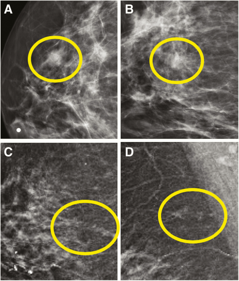
Figure 5.
Examples of low-grade DCIS presenting mammographically. In the first example (A and B), a round mass with obscured margins (circles) was identified in the upper outer quadrant of the right breast at anterior depth on 2D screening mammogram views (BB marker denotes the nipple on the craniocaudal view). Pathological evaluation of this mass yielded low-grade DCIS without comedonecrosis arising in association with an intraductal papilloma. In the second example (C and D), a developing focal asymmetry in the right breast was identified on synthetic 2D screening mammogram views in the right breast at 12 o’clock, posterior depth (circles). Pathology at this site revealed low-grade DCIS without comedonecrosis.

