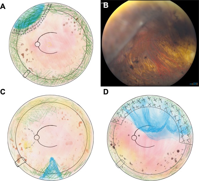
Figure 3.
Surgical drawings of the fundus. Drawings of the fundus were made at the time of diagnosis of retinal detachments. The proband (II:1) had a retinal detachment in the far peripheral region of the superior nasal quadrant OS (A), which is confirmed with fundus photo of the detachment (B). The mother of the proband (I:2) had a far peripheral inferior retinal detachment (C). The sibling of the proband (II:2) had a bullous superior retinal detachment in OS (D).

