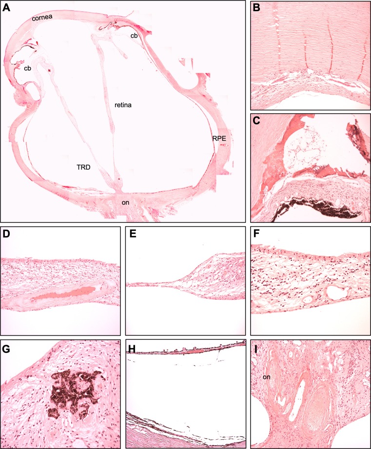
Figure 4.
Histopathology. The OD of the sibling (II:2) was aphakic, phthisical, and showed a complete tractional retinal detachment (TRD) at the time of enucleation (A). The central cornea demonstrated absence of Descemet's membrane along with diffuse endothelial cell loss with secondary stromal edema (B). Neovascularization of the angle with subsequent closure and flattening of the anterior chamber was observed (C). The retinal vasculature demonstrated hyalinization (D). There was thinning and atrophy of the retina (E), cystoid spaces, and subretinal ossification (E, F) along with pigmentary migration (G). The RPE-choroid was thin (H). Severe optic nerve atrophy was observed (I).

