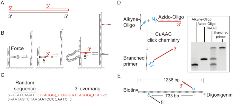
Figure 1.
Rescue-rope-strategy for examining an artificial telomere with an open end. (A) Telomere DNA forms a triplex conformation. Red and black indicate the G-rich and C-rich strand, respectively. (B) Rescue-rope-strategy for probing a telomere with a free end. Conventional mechanical pulling assays can repetitively manipulate structures with a closed end, e.g. a DNA hairpin (left). A structure with a free and open end cannot undergo circles of mechanical pulling-relaxing (middle). A rescue-rope-strategy assisted by dsDNA allows manipulations repetitively on a structure with a free end, e.g. a telomere (right). Color coding the same as (A). (C) The design of an artificial telomere. A random sequence at the blunt upstream end assures the same configuration of the artificial telomere in melting/reannealing circles. The number of TTAGGG motif can vary in a duplex and 3′ overhang. Here, n = 5 and m = 2. (D) Click chemistry to generate a branched primer using alkyne-oligo and azido-oligo. The gel result of polyacrylamide gel electrophoresis shows the reactants, products of Cu(I)-catalyzed azide-alkyne cycloaddition (CuAAC) and branched primer carrying a telomeric G-rich strand after purification. (E) Final construct of an artificial telomere on a rescue-rope DNA. The telomeric G-rich (red) and C-rich (black) ssDNA are positioned at two sites 733 bp away on the rescue-rope DNA, which carries biotin and digoxigenin modifications.

