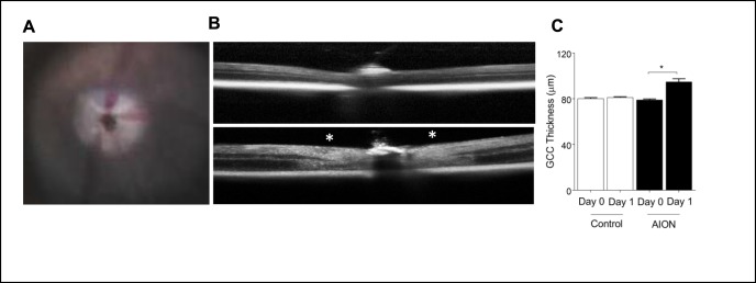
Figure 1.
Photochemical thrombosis model of AION. (A) Mouse fundus photography immediately after AION showing whitening of optic nerve head due to ischemia. (B) Representative images of OCT line scan through optic disc of control (top) and AION (bottom) eyes 1 day after AION. Swelling around the optic nerve head is indicated by white asterisks. (C) Bar graph showing OCT GCC thickness measurements in control and AION eyes at day 0 and 1 (n = 6, *P = 0.0001, 1-way ANOVA with Tukey multiple comparisons test). AION, anterior ischemic optic neuropathy; GCC, ganglion cell complex; OCT, optical coherence tomography.

