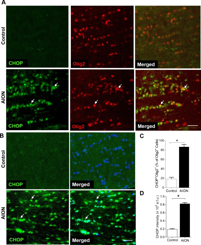
Figure 5.
Significant increase of the percentage of CHOP+Olig2+ cells and intensity of CHOP immunoreactivity in the anterior optic nerve 1 day after AION. (A) Increase in CHOP+Olig2+ oligodendrocytes in the optic nerve 1 day after AION. (B) Representative images and (C) bar graph showing significantly increased number of CHOP+DAPI+ cells in the optic nerve 1 day after AION (*P = 0.03, Mann-Whitney U test). (D) Bar graph showing significantly increased CHOP immunoreactivity in the optic nerve 1 day after AION (*P = 0.03, Mann-Whitney U test). Scale bar: 25 μm.

