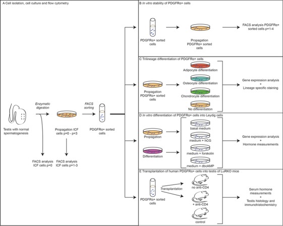
Figure 1.

Overview of the methods performed on ICF and PDGFRα + cells. From a testicular tissue sample, interstitial cells were isolated, propagated and sorted for HLA+/CD34−/PDGFRα+. During propagation of ICF cells, flow cytometry analyses were performed at every passage (A). After sorting, the cells were used to study stability of PDGFRα expression during propagation in culture (B), trilineage differentiation potential (C), in vitro differentiation into Leydig cells (D) and in vivo differentiation into Leydig cells after transplantation into testis of LuRKO mice (E). ICF: interstitial cell fraction, PDGFRα: platelet-derived growth factor receptor alpha; LuRKO: luteinizing hormone receptor knock-out.
