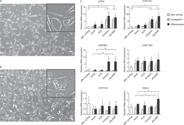
Figure 4.

In vitro differentiation into Leydig cells. After 4 weeks of culture, the morphology of the HLA-A,B,C+/CD34−/PDGFRα+ cells differ when cultured in propagation medium (A) or when cultured in differentiation conditions (B). Scale bar is 100 μm. RNA expression analyses for Leydig cell-specific enzymes and products STAR, CYP11A1, HSD3B2, HSD17B3, CYP17A1 and INSL3 (C). Data are expressed as mean ± SEM (n = 4). Letters a and b indicate significant difference in mRNA expression between propagation and differentiation culture conditions using a Tukey post-hoc test (P < 0.05). The asterisk in the graphs (*) indicates a significant difference in mRNA expression based on 24-h stimulation conditions using a Dunnet post-hoc test (P < 0.05). STAR: steroidogenic acute regulatory protein; CYP11A1: cytochrome p450 family 11 subfamily A member 1; HSD3B2: 3β-hydroxysteroid dehydrogenase type II; HSD17B3: 17β-hydroxysteroid dehydrogenase type III; CYP17A1: cytochrome p450 family 17 subfamily A member 1; INSL3: insulin like 3.
