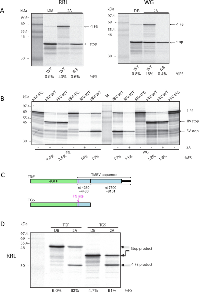
Figure 2.
2A stimulation of PRF. (A) RNAs containing the TMEV PRF signal were translated in RRL (left) or WG (right) with 1.8 μM recombinant 2A (lanes 3–4) or with dialysis buffer (DB). SS indicates a shift site mutant. Products generated by ribosomes that do not frameshift (stop) or that enter the −1 reading frame (−1 FS) are indicated. Markers are in lane 1. (B) RNAs containing the IBV or HIV PRF signals were translated in WG or RRL with or without 1.8 μM recombinant TMEV 2A protein. IFC indicates in-frame controls showing the position at which the frameshift products migrate. Markers are in the middle lane. (C) Schematic of eGFP-based constructs TGF and TG5. TGF (‘TMEV GFP full’) contains the TMEV shift site (G_GUU_UUU) preceded by 14 nt 5′ and followed by 186 nt 3′, fused to the last 480 nt of the TMEV polyprotein ORF, the entire TMEV 3′ UTR and 21 nt of poly(A). TG5 (‘TMEV GFP 5′’) is similar but lacks the 480 nt + 3′ UTR + poly(A) region. (D) TGF and TG5 RNAs were translated in RRL with 1.8 μM recombinant 2A (lanes 3 and 5) or with dialysis buffer (DB; lanes 2 and 4). Markers are in lane 1.

