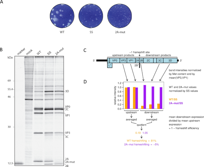
Figure 9.
Mutating 2A inhibits frameshifting in TMEV. (A) Plaque morphology of WT, SS and 2A-mut viruses on BHK-21 cells. (B) Metabolic labelling of BHK-21 cells mock-infected or infected with WT, SS or 2A-mut viruses. Positions of TMEV proteins are indicated. (C) Schematic of the TMEV genome. UTRs are indicated in black and CDSs in pale blue. The lengthy 5′ UTR contains an IRES that directs translation of the polyprotein ORF (L-VP0-VP3-VP1-2A-2B-2C-3A-3B-3C-3D), its frameshift truncation (L-VP0-VP3-VP1-2A-2B*), and the overlapping L* ORF. The tiny 2B* protein shares its N-terminal 6 aa with 2B, whereas its C-terminal 8 aa are encoded in the −1 reading frame. (D) Ratio of band intensities between WT and SS, or 2A-mut and SS viruses.

