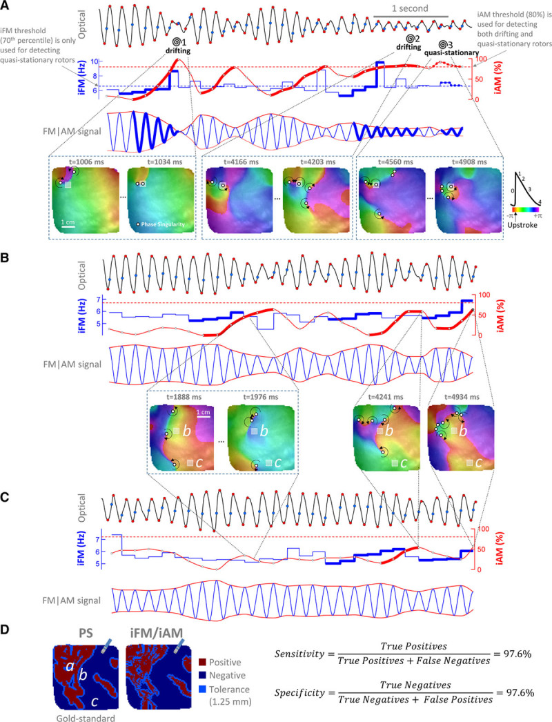
Figure 2.

Examples of instantaneous frequency modulation (iFM)/instantaneous amplitude modulation (iAM) from an optical movie in a persistent atrial fibrillation (PersAF) sheep heart (Online Movie I). A, Top, signal from a pixel (gray square in bottom maps, enlarged to ease visualization) crossed by a phase singularity (PS) in a figure-of-eight reentry. Spirals mark the times at which a PS (white circles) passes by the pixel. Blue points, activation times. Red points, start and end of phase-0. Activation times and phase-0 amplitude excursions are used to generate the iFM (second row, in blue) and the iAM (second row, in red) signals, respectively. Time intervals with sustained simultaneously increasing iFM (second row, thick blue tracings) and iAM (thick red tracings) reaching a prespecified iAM threshold (horizontal red dashed line) are detected. Afterward, the rotor is still considered to be around while iAM keeps over the threshold regardless of iFM (see rotor no. 2 and Online Figure/Movie II). Time intervals with simultaneously high, but not necessarily increasing, iFM and iAM above prespecified thresholds (horizontal blue and red dashed lines, respectively) are detected as quasistationary rotors (see rotor no. 3). The third row displays a synthetic FM | AM signal in which the rotational-footprint positive intervals are highlighted. (Continued )Figure 2 Continued. B, Signal from a pixel close to areas swept by drifting rotors but not actually crossed by their associated PS. Note that there are still intervals with simultaneously increasing iFM and iAM. However, iAM does not reach the prespecified threshold. Therefore, the algorithm properly classifies the pixel as rotational-footprint negative. C, Signal from a pixel far from areas swept by drifting rotors. Increasing iAM, although still present, is not as noticeable as in pixels very close to or actually crossed by drifting rotors. Therefore, the algorithm also classifies this pixel as rotational-footprint negative. D, Comparison between the pixels actually crossed by a PS (PS map, “gold standard”) and the pixels detected by the single-signal algorithm as rotational-footprint positive (iFM/iAM map). The size of a conventional ablation catheter is shown for reference. The signals shown in A–C were taken from pixels a–c. Additional examples are shown in Online Figure IV.
