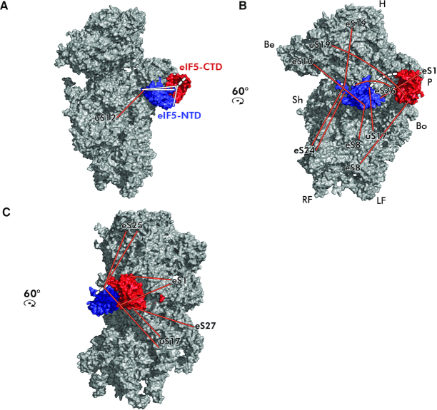
Figure 9.
Placement of both terminal domains of eIF5 on the 40S ribosomal subunit modeled based on the obtained cross-links. (A–C) The N-terminal (PDB code: 2E9H—in blue) and the C-terminal (PDB code: 2FUL—in red) domains of eIF5 were modeled onto the intersubunit side of the 40S subunit (PDB code: 3JAM) based on the obtained cross-links. The linker region, unresolved in the available 3D structures, is depicted as a dashed white line. Cross-links between ribosomal proteins and eIF5 are depicted in orange, intramolecular cross-links between the eIF5 halves in white; identities of small ribosomal proteins are indicated at the end of each cross-link. For details see the main text.

