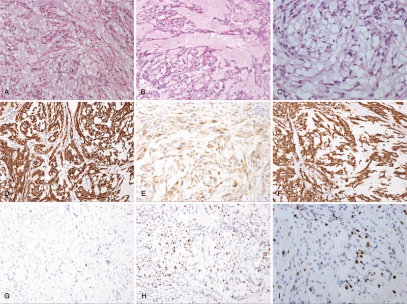
Figure 8.

Pathologic histology of spinal tumors. (A–D) Microphotography showing characteristic nests of tumor cells separated by vascular septa (Zellballen) with cells showing significant nuclear pleomorphism with prominent nucleoli (H&E, original magnification 100×, 100×, 100×, and 200×). (E) AE1/AE3 immunostaining is strongly positive in the epithelial cells. (F) EMA immunostaining shows positive staining in the tumor cells. (G) Vimentin immunostaining shows positive staining in the tumor cells. (H, I) Ki-67 immunostaining shows 15% Ki-67 positive cells. Ki-67 staining is localized in the tumor nuclei.
