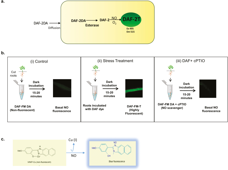
Fig. 1.
In vivo NO fluorescence visualized by using DAF fluorescent dyes. (a) Reaction mechanisms of DAF-2DA. The diacetate groups of DAF dyes are removed by intracellular esterases, the diffused DAF-2 in the presence of NO (and O2) forms highly fluorescent DAF-2T. (b) Suggested use of DAF dyes for measurement of NO under stress conditions. (i) Shows DAF fluorescence in roots of control seedlings incubated in DAF-FM-DA (10 μM in 50 mM HEPES buffer, pH 7.2) for 15 min in the dark. (ii) NO production under stress conditions; after incubation, roots shows intense DAF-FM fluorescence. (iii) Similarly, roots from stress-induced plants display basal NO fluorescence when cPTIO (an NO scavenger) is used along with DAF-FM under the same incubating conditions. (c) MNIP-Cu probe reacts directly with NO and gives blue fluorescence at 420 nm.

