Figure 3.

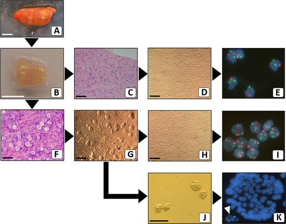
Flow scheme for separating ovarian cortex cellular constituents prior to FISH analysis. After the surgical removal of the intact ovary (A), cortical fragments were prepared. Part of one representative cortex fragment (B) was analysed by standard haematoxylin–eosin staining for the presence of follicles. When no follicles were present (C), the remaining part of the fragment was used to make a suspension of stromal cells (D), for interphase FISH with chromosome X (green) and chromosome 18 (red)-specific probes (E). When follicles were present (F), the remaining part of the cortex fragment was used to make a cell suspension (G) from which small follicles were manually picked up. These isolated follicles were subjected to further digestion (J) and subsequently analysed by FISH. The white arrowhead points to the signals from the oocyte (K). Part of the remaining cell suspension (H) was used for FISH analysis of stromal cells (I). Bars represent 1 cm (A and B) or 100 μm (C, D, F–H and J). Original magnification of FISH signals was ×630 (E, I and K).
