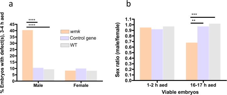
Fig 4. wmk-induced embryonic defects are enriched in males.
Data are from pooled embryos (both sexes, expressing and non-expressing, see methods) with either wmk, the control gene, or a WT background. (A) Graph quantitating the percentage of 3–4 h AED embryos (males or females) that have at least one defect (wmk males N = 228, control gene males N = 190, WT males N = 170, wmk females N = 240, control gene females N = 200, WT females N = 158). (B) Graph quantitating the sex ratio of viable embryos (not degraded, no visible defects) across two development times (1–2 h wmk, N = 105 m, 111 f; 1–2 h control gene, N = 30 m, 141 f; 1–2 h WT, N = 112 m, 115 f; 16–17 h wmk, N = 104 m, 154 f; 16–17 h control gene, N = 116 m, 120 f; 16017 h WT, N = 110 m, 108 f). m = male, f = female. Statistics were performed with a Chi-square test comparing the three genotypes within each category (male or female in (A) and 1–2 h or 16–17 h in (B)). These experiments were performed once. ** p<0.01, *** p<0.001, ****p<0.0001.

