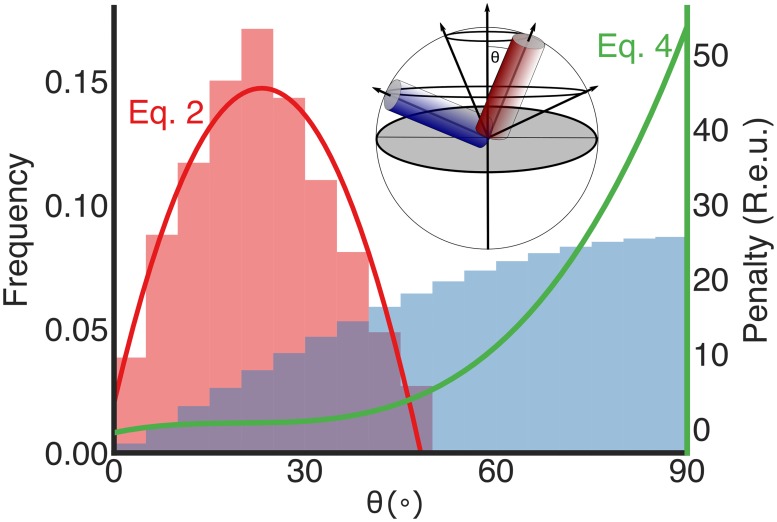
Fig 2. Observed versus expected tilt angles in membrane-spanning helices relative to the membrane normal.
The distribution of helix tilt angles (θ in the inset sphere) in natural membrane proteins shows a strong preference for small angles (red bars, left), whereas the distribution resulting from random conformational sampling is proportional to sin(θ) (blue bars) [30] significantly overrepresenting large tilt angles. The MPSpanAngle energy term (green line; Eq 4) penalises large tilt angles and focuses ab initio conformational sampling on tilt angles observed in membrane-protein structures. inset The expected distribution of helix-tilt angles is proportional to the circumference of a circle plotted by that helix around an axis perpendicular to the membrane-normal (panel adapted from ref. [30]). The membrane plane is depicted as a grey circle.

