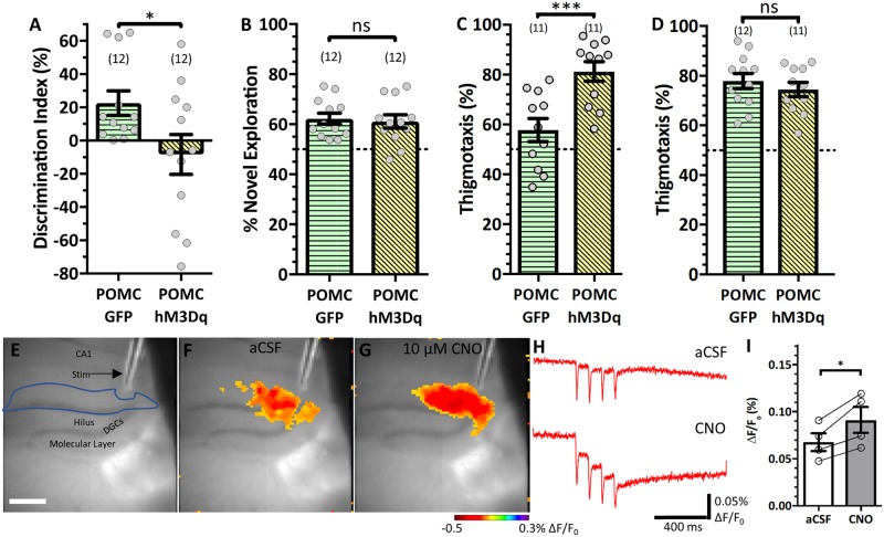
Figure 8.
Selectively activating DGCs compromises SOR. POMC-Cre mice were injected with either GFP or hM3Dq and received 0.3 mg/kg CNO 1 h prior to test trials throughout behavioural testing. (A) SOR performance [t(22) = 2.196, P = 0.039]. (B) Novel object recognition performance [t(22) = 0.3256, P = 0.748]. (C) Open field anxiety analysis [t(20) = 3.857, P = 0.001]. (D) SOR test trial anxiety analysis [t(20) = 0.8083, P = 0.428]. (E) Representative micrograph of voltage sensitive dye recording configuration in an hM3Dq-expressing slice. Scale bar = 425 µm. (F and G) ΔF/F0 (%) max response in artificial CSF (aCSF) (F) and 10 µM CNO (G). (H) Representative traces of ΔF/F0 in artificial CSF (top) and CNO (bottom). (I) Quantification of response to stimulation in the DGC+molecular layer [t(3) = 4.302, P = 0.023]; each circle represents the average of two to three slices per animal. Statistical comparisons were Student’s t-tests (A–D) or two-tailed paired t-tests (I). Data are represented as mean ± SEM. Each circle represents an animal (number of animals indicated in parentheses). nsP > 0.05, *P < 0.05, **P < 0.01.

