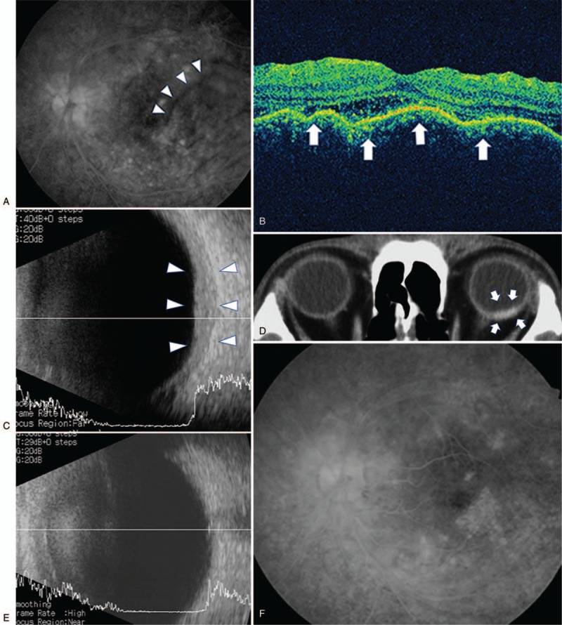
Figure 1.

(A) Fluorescein angiography of the left eye showing peripheral vasculitis characteristic of Behcet disease and retinal folds at the posterior pole. (B) Optical coherence tomography showing choroidal folds (arrows) in the left eye. (C) B-scan ultrasonography of the left eye demonstrating thickened sclera (arrowheads). (D) CT scan of the patient showing contrast enhancement (arrows) localized to the posterior sclera of the left eye. (E) B-scan ultrasonography of the left eye 43 weeks after treatment showing disappearance of choroid hypertrophy. (F) Fluorescein angiography of the left eye 43 weeks after treatment showing disappearance of retinal folds.
