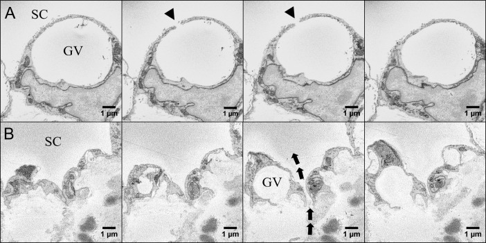
Figure 3.
Serial block-face scanning electron micrographs of GV, intracellular pore (I-pore), and border pore (B-pore) in the IW endothelium of SC. (A) Serial images of an IW endothelial cell with a GV and an I-pore (arrowhead) leading into SC. (B) Serial images of a B-pore (arrows) located at the border between two adjacent inner wall cells of SC.

