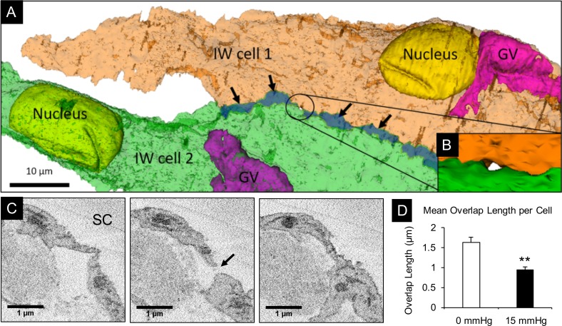
Figure 11.
Analysis of B-pores and overlapping cell margins between two IW cells. (A) Two adjacent IW cells (green and orange) were made semitransparent to show overlapping cell margins (dark green region indicated by arrows) and a B-pore (encircled). (B) Higher magnification snapshot of reconstructed pore encircled in (A). (C) Three serial block-face scanning electron micrographs from which the B-pore (arrow) was identified and reconstructed. (D) Comparison of the mean overlap length (OL) in IW cells from immersion-fixed eyes (n = 12 cells) and perfusion-fixed eyes (n = 12 cells). Mean OL was significantly less in perfusion-fixed eyes (0.93 ± 0.24 μm) compared to immersion-fixed eyes (1.63 ± 0.12 μm; P < 0.01). Error bars: SEM. **P < 0.01.

