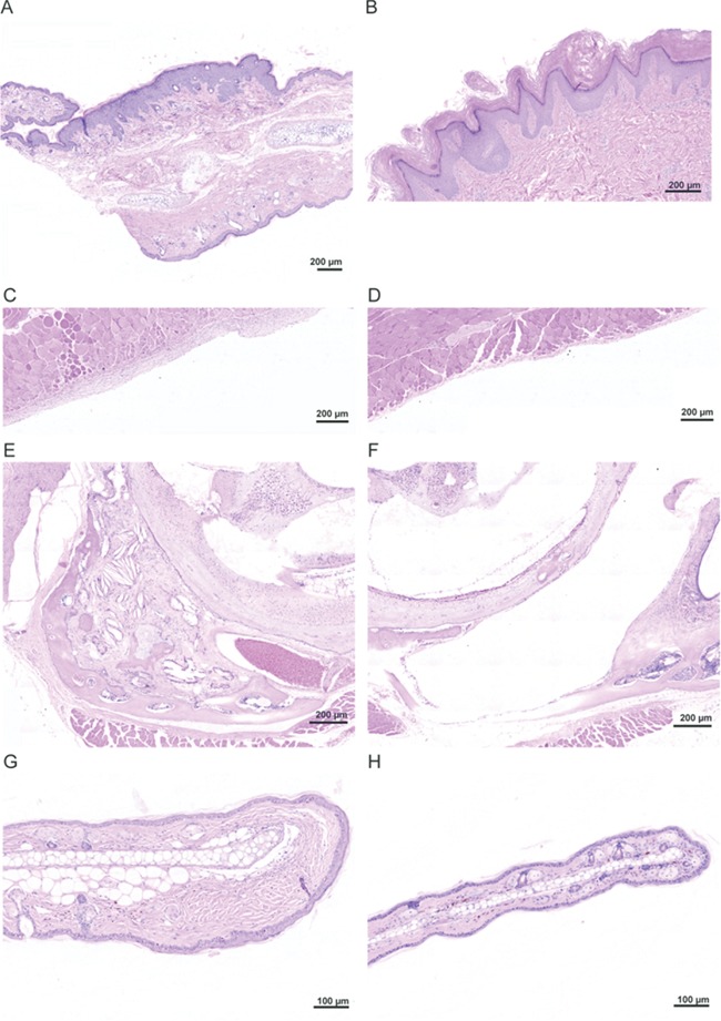
Figure 6.

Hyperplasia and stromal expansion in Proteus syndrome mice. (A) A region of epidermal hyperplasia on the ear of chimera E-2. The upper edge showed expanded epidermis whereas the lower edge epidermis was normal thickness. (B) A linear epidermal nevus from a patient with Proteus syndrome. Note the similarity to the epidermal hyperplasia shown in chimera E-2 shown in panel A. (C) Expansion of the stroma lining the peritoneal wall in chimera E-2. (D) Normal thickness of stroma in an adjacent region of the peritoneal wall of this animal. (E) Overgrowth of multiple tissues in the left middle ear of chimera A-9. Expanded stroma, osseous metaplasia, cholesterol clefts, and several small cysts and areas of mineralization were seen. (F) Normal right middle ear of that same animal for comparison. (G) Increased collagen in the dermis at the tip of the ear in chimera E-4. (H) A normal ear from control T2C-81 for comparison.
