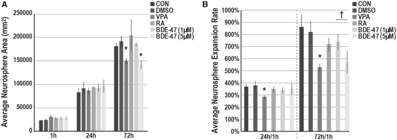
Figure 4.
BDE-47 exposures and neurosphere expansion. (A) Average neurosphere size (µm2) in cultures exposed to BDE-47, 0.1% DMSO (VEH) or media only (CON). Suspended neurospheres (preexposed for 3 days) were plated on Cellstart. At 1, 24, and 72 h, successive phase images of single neurospheres (x—= 12.1 neurospheres per group; minimum ≥ 5) were acquired. Total neurosphere area was quantified (ImageJ) and averaged across experiments (n = 4; 1 and 24 h; n = 3; 72 h). B, Average neurosphere expansion rates at 24 or 72 h in comparison with 1 h measurements. In each experiment, expansion rate was determined, and then, averaged across experiments. Significant differences (p < .05) due to exposure as compared with the vehicle control are indicated by double-crosses (‡, ANOVA) or asterisks (t test).

