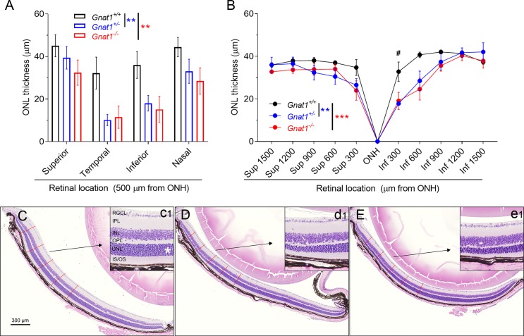
Figure 1.
Homozygous and heterozygous knockouts of rod-transducin (Gnat1) slightly increase susceptibility to light damage in mice. (A) ONL (i.e., photoreceptor nuclei layer) thickness analysis from OCT images taken at 7 days after BLE shows that Gnat1+/− (blue bars, n = 8) and Gnat1−/− (red bars, n = 5) have thinned ONL when compared with Gnat1+/+ (black bars, n = 7) mice. Geisser-Greenhouse corrected RM ANOVA was used followed by Bonferroni's post hoc tests to compare between-subjects main effect: Gnat1+/+ vs. Gnat1+/− and Gnat1+/+ vs. Gnat1−/−. Significance denoted by **P < 0.01. (B) Morphometry analysis from histological sections shows that central inferior (Inf) retina is thinned both in Gnat1+/− (n = 7) and Gnat1−/− (n = 5) mice in relation to the Gnat1+/+ (n = 6) mouse, but superior (Sup) retina is relatively spared. Geisser-Greenhouse corrected RM ANOVA was used followed by Bonferroni's post hoc tests to compare between-subjects main effect: Gnat1+/+ vs. Gnat1+/− and Gnat1+/+ vs. Gnat1−/−. Significance denoted by **P < 0.01 and ***P < 0.001. Post hoc test to compare the genotypes in different locations: #P < 0.05. (C) A representative sample of H&E stained retinal cross-section from a Gnat1+/+ mouse. Inferior retina is shown at ONH level in relation to temporal-nasal orientation. Red lines illustrate the measurement points of ONL thickness used for statistical analysis in image B. The inset (c1) shows a magnification image between 600 and 900 μm from ONH, and the asterisk marks the ONL. (D, E) Similar images as C, but from Gnat1+/− (D and inset d1) and Gnat1−/− (E and inset e1) mice. RGCL, retinal ganglion cell layer; IPL, inner plexiform layer; INL, inner nuclear layer; IS, photoreceptor inner segments; OS, photoreceptor outer segments.

