It is a pleasure to join the other authors in this issue to honor Dr Arnold Levine and the remarkable impact of his research. Our paths crossed with Arnie’s when our genetic analyses of colorectal cancers led us to investigate the p53 gene and identify it as a tumor suppressor gene. p53 was initially identified by Arnie and others as an oncogene and tumor antigen, nearly a decade before its role as a tumor suppressor gene was revealed. We fondly remember Arnie visiting our lab, and we visiting his, to discuss the implications of the genetic alterations our group identified in human cancers and how we could work together to investigate the voluminous new questions these mutations raised.
The rationale for the existence of tumor suppressor genes
The hunt for tumor suppressor genes was a high-risk endeavor in the 1980s. Somatic cell hybridization studies provided early evidence of the existence of tumor suppressor genes by showing that tumorigenic growth was a recessive trait. Hybrid cell fusions of malignant cells with non-malignant cells could suppress tumorigenic growth, and microcell mediated-transfer of specific human chromosomes could suppress the tumorigenic growth of human cancer cell lines in immunodeficient mice (Harris et al., 1969; Klinger, 1982; Peehl and Stanbridge, 1982; Harris, 1988). Alfred Knudson’s insightful analyses of retinoblastoma incidence connected the concept of tumor suppressor genes with human disease pathogenesis (Knudson, 1971, 1985). What later became known as the ‘two-hit hypothesis’ proposed that patients with familial retinoblastoma inherited the first ‘hit’, an inactivating mutation in one allele of a tumor suppressor gene that predisposed them to the disease. A somatic mutation, or second ‘hit’, in the remaining allele would result in complete loss of function of the gene and contribute to tumorigenesis. In sporadic tumors, both hits were hypothesized to occur within the same somatic cell. Due to the much lower likelihood of two somatic mutations, sporadic retinoblastomas were typically unilateral and arose at a later age.
Cytogenetic abnormalities and submicroscopic deletions of chromosome 13q14 revealed the chromosomal location of the putative tumor suppressor gene in retinoblastoma (Francke and Kung, 1976; Knudson et al., 1976; Benedict et al., 1983; Cavenee et al., 1983, 1985; Godbout et al., 1983). These studies later led to the positional cloning of the RB1 gene, thus validating the two-hit hypothesis (Friend et al., 1986; Fung et al., 1987; Lee et al., 1987).
Mapping the chromosome 17p tumor suppressor locus
In the late 1980s, our group mapped regions of chromosomal loss in colorectal cancer to identify the locations of tumor suppressor genes. The highest frequency loss involved the short arm of chromosome 17 (17p), which occurred in >75% of colorectal carcinomas (Reichmann et al., 1981; Muleris et al., 1985; Fearon et al., 1987; Vogelstein et al., 1988). Sporadic colorectal cancers posed a significant challenge compared to hereditary cancer predisposition syndromes like retinoblastoma where small constitutional deletions narrowed down the target area for analysis. To more precisely localize the candidate tumor suppressor, we performed Southern blots with a panel of 20 different polymorphic markers on 17p to evaluate loss of heterozygosity (LOH) in 58 paired samples of colorectal carcinoma and matched normal colorectal tissues. This analysis identified a minimal common region of deletion shared among all tumors in which any LOH was observed. This region encompassed approximately half of 17p (Baker et al., 1989). With today’s genomic maps, we can estimate that the common region of deletion spanned >12.5 megabase pairs of DNA and contained ~577 genes, including 480 protein-coding genes. Relative to today, genomic maps in 1988 were extremely sparse and much of the genome could be considered uncharted territory in terms of the density and identity of genes. Identifying the tumor suppressor gene within this area was a daunting prospect, and we did not consider it likely when we started this project in the mid-80’s that the gene could actually be identified within a time-frame consistent with a pre-doctoral thesis. Remember that at the time (1985), oncogenes were already known but tumor suppressor genes were mythical beasts, predicted to exist but not yet sighted.
p53: oncogene or tumor suppressor gene
p53 was identified in 1979 by Arnie Levine’s group, as well as four other groups, as a host protein bound to large T antigen in cells infected with the transforming virus SV40 (Kress et al., 1979; Lane and Crawford, 1979; Linzer and Levine, 1979; Melero et al., 1979; Smith et al., 1979), and by another group as a transformation-related antigen in chemically induced mouse sarcomas (DeLeo et al., 1979). Multiple lines of research initially supported the hypothesis that p53 was an oncogene, including association of the p53 protein with viral transforming proteins of SV40 or adenovirus in infected cells, and elevated expression of p53 protein in transformed cells and human tumor cell lines. The p53 cDNA was cloned by several groups, often from tumor cell lines with robust p53 protein expression, and expression of p53 cDNA could cooperate with other oncogenes to transform primary mouse cells and increase tumorigenic growth of established tumor cells. While multiple lines of evidence supported the widespread view that p53 was an oncogene, there were some observations that had been interpreted as suggesting a more complex story (Wolf and Rotter, 1985; Eliyahu et al., 1988; Finlay et al., 1988; Hicks and Mowat, 1988; Hinds et al., 1989). One explanation for these complexities was that p53 was actually a tumor suppressor gene rather than an oncogene. This interpretation was largely based on the fact that the normal p53 gene from mice inhibited cell growth. The investigators were appropriately cautious about this interpretation; we now know that many oncogenes, such as BCL2 and IDH1, when overexpressed, can inhibit growth (Vogelstein et al., 2013).
Testing the two-hit hypothesis
We began our studies of p53 in 1987, assuming that it was an oncogene, for the reasons described above. In particular, we initially did not think that it was the tumor suppressor gene target of the chromosome 17p deletions for which we were searching. But because the p53 gene was in the middle of the ‘lost chromosome region’ on chromosome 17p in colorectal cancers, and because it had been implicated to play a role in neoplasia, we thought we had to eliminate it as a candidate before continuing the search for the actual tumor suppressor gene on chromosome 17p. Based on the two-hit hypothesis, we began our evaluation by looking for evidence of homozygous deletion or rearrangements of the p53 locus in colorectal carcinomas; none were detected by Southern blot analysis. Northern blot analysis of colorectal carcinomas showed that p53 was expressed in most colorectal carcinomas, and there was no evidence of abnormally sized transcripts. To test for more subtle alterations in p53, we selected a single tumor that had an allelic deletion of chromosome 17p and sequenced the remaining p53 allele in this tumor (Baker et al., 1989). To our initial disbelief, it contained a p53 missense mutation in the remaining allele at codon 143 (V143A). This change was not present in the normal colon mucosa of this patient, so it was somatic, in accordance with the two-hit hypothesis. But a change from valine to alanine did not necessarily mean that the encoded protein would be drastically different, and we wished to confirm this observation in a second colorectal cancer in which one chromosome 17p allele was lost. The second tumor did indeed contain a p53 mutation, this time at codon 175 (R175H).
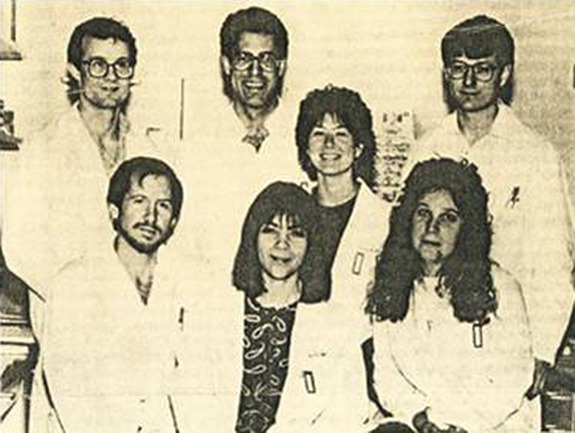
Finally, we showed that these types of genetic alterations were not confined to colorectal cancers. Working with Janice Nigro and others in our lab (Figure 1), we found that 17p allelic losses coupled with missense mutations in the remaining p53 allele occurred in many tumors of the brain, breast, lung, and mesenchyme, in addition to a larger cohort of colorectal cancers (Nigro et al., 1989; Baker et al., 1990). These studies also showed that there were ‘hotspots’ in p53—mutations that occurred much more frequently in some positions than others—and that the mutations occurred relatively late in tumorigenesis, perhaps driving the transition from benign tumors (adenomas) to malignant ones (carcinomas).
Figure 1.

Members of the Vogelstein Lab, circa 1989. Front row: Bert Vogelstein, Janice M. Nigro, Ann C. Preisinger; middle row: Suzanne J. Baker; back row: J. Michael Ruppert, Eric R. Fearon, Kenneth W. Kinzler.
p53 today
By satisfying the two-hit hypothesis, our initial studies provided definitive genetic evidence that p53 was a tumor suppressor gene. It is interesting that even today, functional studies cannot reliably distinguish between tumor suppressor genes and oncogenes, the only definitive way to classify a cancer driver gene is through genetic approaches (Vogelstein et al., 2013). And now that extensive genome-wide sequencing of human cancers has provided an unbiased view of the mutation landscape, we know that p53 is the most commonly mutated gene in human cancer. With >28000 mutations reported (p53.IARC.fr), missense mutation remains the most common mechanism for p53 inactivation, accounting for almost 75% of all p53 mutations. We hope that Arnie, on the occasion of his 80th birthday, can derive considerable satisfaction from the fact that his work inspired a revolution in the understanding of human cancer.
[We apologize to the many scientists who have made important contributions to p53 research that could not be cited in this brief retrospective.]
References
- Baker S.J., Fearon E.R., Nigro J.M., et al. (1989). Chromosome 17 deletions and p53 gene mutations in colorectal carcinomas. Science 244, 217–221. [DOI] [PubMed] [Google Scholar]
- Baker S.J., Preisinger A.C., Jessup J.M., et al. (1990). p53 gene mutations occur in combination with 17p allelic deletions as late events in colorectal tumorigenesis. Cancer Res. 50, 7717–7722. [PubMed] [Google Scholar]
- Benedict W.F., Murphree A.L., Banerjee A., et al. (1983). Patient with 13 chromosome deletion: evidence that the retinoblastoma gene is a recessive cancer gene. Science 219, 973–975. [DOI] [PubMed] [Google Scholar]
- Cavenee W.K., Dryja T.P., Phillips R.A., et al. (1983). Expression of recessive alleles by chromosomal mechanisms in retinoblastoma. Nature 305, 779–784. [DOI] [PubMed] [Google Scholar]
- Cavenee W.K., Hansen M.F., Nordenskjold M., et al. (1985). Genetic origin of mutations predisposing to retinoblastoma. Science 228, 501–503. [DOI] [PubMed] [Google Scholar]
- DeLeo A.B., Jay G., Appella E., et al. (1979). Detection of a transformation-related antigen in chemically induced sarcomas and other transformed cells of the mouse. Proc. Natl Acad. Sci. USA 76, 2420–2424. [DOI] [PMC free article] [PubMed] [Google Scholar]
- Eliyahu D., Goldfinger N., Pinhasi-Kimhi O., et al. (1988). Meth a fibrosarcoma cells express two transforming mutant p53 species. Oncogene 3, 313–321. [PubMed] [Google Scholar]
- Fearon E.R., Hamilton S.R., and Vogelstein B. (1987). Clonal analysis of human colorectal tumors. Science 238, 193–197. [DOI] [PubMed] [Google Scholar]
- Finlay C.A., Hinds P.W., Tan T.H., et al. (1988). Activating mutations for transformation by p53 produce a gene product that forms an hsc70-p53 complex with an altered half-life. Mol. Cell. Biol. 8, 531–539. [DOI] [PMC free article] [PubMed] [Google Scholar]
- Francke U., and Kung F. (1976). Sporadic bilateral retinoblastoma and 13q- chromosomal deletion. Med. Pediatr. Oncol. 2, 379–385. [DOI] [PubMed] [Google Scholar]
- Friend S.H., Bernards R., Rogelj S., et al. (1986). A human DNA segment with properties of the gene that predisposes to retinoblastoma and osteosarcoma. Nature 323, 643–646. [DOI] [PubMed] [Google Scholar]
- Fung Y.K., Murphree A.L., T'Ang A., et al. (1987). Structural evidence for the authenticity of the human retinoblastoma gene. Science 236, 1657–1661. [DOI] [PubMed] [Google Scholar]
- Godbout R., Dryja T.P., Squire J., et al. (1983). Somatic inactivation of genes on chromosome 13 is a common event in retinoblastoma. Nature 304, 451–453. [DOI] [PubMed] [Google Scholar]
- Harris H. (1988). The analysis of malignancy by cell fusion: the position in 1988. Cancer Res. 48, 3302–3306. [PubMed] [Google Scholar]
- Harris H., Miller O.J., Klein G., et al. (1969). Suppression of malignancy by cell fusion. Nature 223, 363–368. [DOI] [PubMed] [Google Scholar]
- Hicks G.G., and Mowat M. (1988). Integration of Friend murine leukemia virus into both alleles of the p53 oncogene in an erythroleukemic cell line. J. Virol. 62, 4752–4755. [DOI] [PMC free article] [PubMed] [Google Scholar]
- Hinds P., Finlay C., and Levine A.J. (1989). Mutation is required to activate the p53 gene for cooperation with the ras oncogene and transformation. J. Virol. 63, 739–746. [DOI] [PMC free article] [PubMed] [Google Scholar]
- Klinger H.P. (1982). Suppression of tumorigenicity. Cytogenet. Cell Genet. 32, 68–84. [DOI] [PubMed] [Google Scholar]
- Knudson A.G. Jr, Meadows A.T., Nichols W.W., et al. (1976). Chromosomal deletion and retinoblastoma. N. Engl. J. Med. 295, 1120–1123. [DOI] [PubMed] [Google Scholar]
- Knudson A.G., Jr. (1971). Mutation and cancer: statistical study of retinoblastoma. Proc. Natl Acad. Sci. USA 68, 820–823. [DOI] [PMC free article] [PubMed] [Google Scholar]
- Knudson A.G., Jr. (1985). Hereditary cancer, oncogenes, and antioncogenes. Cancer Res. 45, 1437–1443. [PubMed] [Google Scholar]
- Kress M., May E., Cassingena R., et al. (1979). Simian virus 40-transformed cells express new species of proteins precipitable by anti-simian virus 40 tumor serum. J. Virol. 31, 472–483. [DOI] [PMC free article] [PubMed] [Google Scholar]
- Lane D.P., and Crawford L.V. (1979). T antigen is bound to a host protein in SV40-transformed cells. Nature 278, 261–263. [DOI] [PubMed] [Google Scholar]
- Lee W.H., Bookstein R., Hong F., et al. (1987). Human retinoblastoma susceptibility gene: cloning, identification, and sequence. Science 235, 1394–1399. [DOI] [PubMed] [Google Scholar]
- Linzer D.I., and Levine A.J. (1979). Characterization of a 54K Dalton cellular SV40 tumor antigen present in SV40-transformed cells and uninfected embryonal carcinoma cells. Cell 17, 43–52. [DOI] [PubMed] [Google Scholar]
- Melero J.A., Stitt D.T., Mangel W.F., et al. (1979). Identification of new polypeptide species (48-55K) immunoprecipitable by antiserum to purified large T antigen and present in SV40-infected and -transformed cells. Virology 93, 466–480. [DOI] [PubMed] [Google Scholar]
- Muleris M., Salmon R.J., Zafrani B., et al. (1985). Consistent deficiencies of chromosome 18 and of the short arm of chromosome 17 in eleven cases of human large bowel cancer: a possible recessive determinism. Ann. Genet. 28, 206–213. [PubMed] [Google Scholar]
- Nigro J.M., Baker S.J., Preisinger A.C., et al. (1989). Mutations in the p53 gene occur in diverse human tumour types. Nature 342, 705–708. [DOI] [PubMed] [Google Scholar]
- Peehl D.M., and Stanbridge E.J. (1982). The role of differentiation in the suppression of tumorigenicity in human cell hybrids. Int. J. Cancer 30, 113–120. [DOI] [PubMed] [Google Scholar]
- Reichmann A., Martin P., and Levin B. (1981). Chromosomal banding patterns in human large bowel cancer. Int. J. Cancer 28, 431–440. [DOI] [PubMed] [Google Scholar]
- Smith A.E., Smith R., and Paucha E. (1979). Characterization of different tumor antigens present in cells transformed by simian virus 40. Cell 18, 335–346. [DOI] [PubMed] [Google Scholar]
- Vogelstein B., Fearon E.R., Hamilton S.R., et al. (1988). Genetic alterations during colorectal-tumor development. N. Engl. J. Med. 319, 525–532. [DOI] [PubMed] [Google Scholar]
- Vogelstein B., Papadopoulos N., Velculescu V.E., et al. (2013). Cancer genome landscapes. Science 339, 1546–1558. [DOI] [PMC free article] [PubMed] [Google Scholar]
- Wolf D., and Rotter V. (1985). Major deletions in the gene encoding the p53 tumor antigen cause lack of p53 expression in HL-60 cells. Proc. Natl Acad. Sci. USA 82, 790–794. [DOI] [PMC free article] [PubMed] [Google Scholar]


