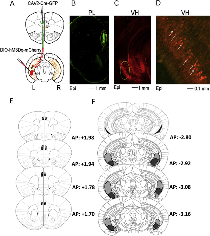
Fig. 2.
Schematic illustrating design, epifluorescent (Epi) patterns of expression, and extent of transfection. A. Schematic of intersectional chemogenetic viral approach. Green circles indicate injection sites of CAV2-Cre-GFP in the PL cortex and red circles injection sites of DREADD-Gq in the VH. Red cells surrounded by the green color represent recombination leading to DREAD-Gq expression. B-C. CAV2-Cre-GFP injection sites in the PL (B) and DREADD-Gq expression in the VH (C). D. Amplification of area surrounded by a yellow dashed line in (B) shows expression restricted to pyramidal ventral cells. E. Extend of diffusion of the CAV-2-cre-GFP virus in PL. F. Extent of transfection in the VH.

