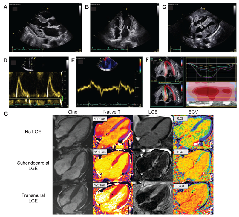
Figure 3.

Typical ECHO and CMR findings in a patient with cardiac amyloidosis. Parasternal longitudinal axis (A) and apical 4-chamber (B) ECHO views show considerably increased LVWT in the absence of ventricular dilation; the myocardial walls appear hyperechogenic. Other characteristic findings include biatrial enlargement and thickening of valve leaflets (A, B) of the interatrial septum (B) and RV free wall (C). A generalized small pericardial effusion is also noticeable (A-C). The profile of LV filling (D) is restrictive, with markedly elevated E wave, reduced A wave, and decreased deceleration time. A decreased E’ wave measurement can be observed on lateral wall tissue Doppler imaging (E). The longitudinal systolic function is impaired with decreased S’ measurement on lateral wall tissue Doppler imaging (E) and markedly reduced longitudinal strain evident on the apical 4-chamber view (F). LV longitudinal strain (F) is preserved at the LV apex but is significantly impaired at the midbasal segments. Each colored curve shows longitudinal strain at 1 of the 6 LV measured segments. Dotted line is the mean. Color map represents the 6 LV segments, with time corresponding to the x-axis. The “bulls-eye” appearance (with apex at the center of the color-coding map) is typical of cardiac amyloidosis. Cardiac magnetic resonance images (G) include 4-chamber cine, corresponding native T1 maps, LGE image with phase-sensitive reconstruction and ECV maps in a patient with no cardiac amyloidosis (upper row) and 2 patients with cardiac amyloidosis (middle row and bottom row). In the upper row, the patient with no cardiac amyloidosis has no LGE and normal native T1 and ECV maps; in the middle row, the patient with cardiac amyloidosis has subendocardial LGE, elevated T1 values and elevated ECV values; in the bottom row, the patient with cardiac amyloidosis has a very high cardiac amyloid load, with transmural LGE, very high native T1 values, and very high ECV values. CMR, cardiac magnetic resonance; ECHO, echocardiography; ECV, extracellular volume; LGE, late gadolinium enhancement; LV, left ventricle; LVWT, left ventricular wall thickness; RV, right ventricle.
