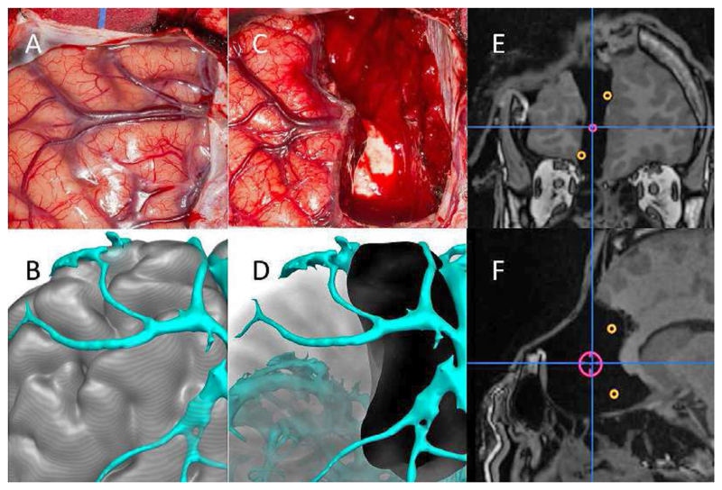
Fig. 2.
Pre- and post-operative recording of resection. A) Intra-operative photograph of left frontal region. B) Modelling of gyral and vascular anatomy. C) Intra-operative photograph of left frontal resection. D) Reconstruction of completed resection. E,F) Coronal and sagittal intra-operative MRI showing completed resection, incorporating neurophysiological landmarks of ictal onset (red) and propagation to anterior cingulum and medial orbito-frontal area (orange). Image courtesy of Mark Nowell.

