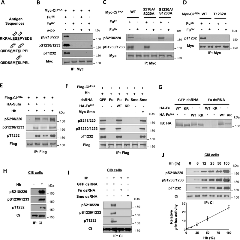
Figure 3. Hh induces Ci phosphorylation at C-I and C-II depending on Smo and Fu kinase activity.
(A) The antigen sequences for generating the three phospho-specific antibodies.
(B-D) Western blot analysis of Myc-Ci−PKA and its variants in S2 cells co-expressed with FuEE and FuGV with the indicated phospho-specific antibodies.
(E) Western blot analysis of Flag-Ci−PKA phosphorylation in S2 cells with or without Sufu coexpression and treated with Hh-conditioned medium or control medium.
(F) Western blot analysis of Flag-Ci−PKA phosphorylation in S2 cells treated with the indicated dsRNA and Hh-conditioned medium and cotransfected without or with the indicated Smo or Fu construct.
(G) Western blot analysis of HA-Fu and HA-FuIns from S2 cells expressing the indicated Fu constructs and treated with GFP or Fu dsRNA.
(H-I) Western blot analysis of endogenous Ci phosphorylation from Cl8 cells treated with or without Hh-conditioned medium and the indicated dsRNA.
(J) Western blot analysis of endogenous Ci phosphorylation (top panels) or ptc-luc reporter assay (bottom panel) in Cl8 cells treated with increasing concentrations of Hh-conditioned medium.

