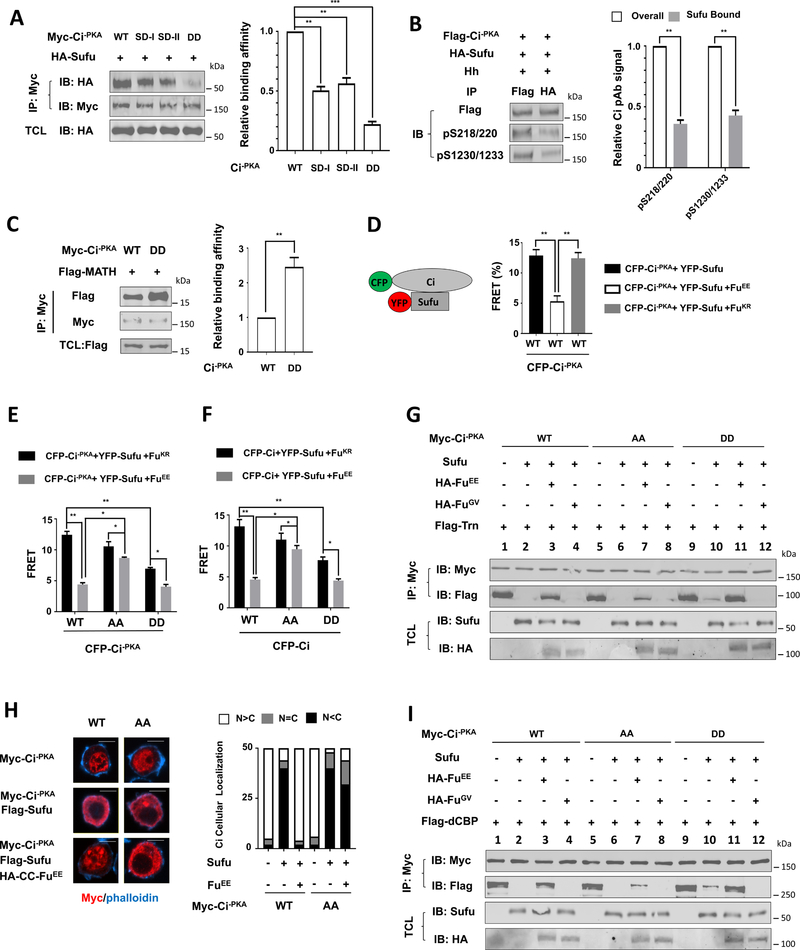
Figure 5. Phosphorylation of Ci reduces its binding to Sufu while increases its binding to Trn and dCBP.
(A-C) Western blot analysis (left panel) and quantitation (right panel) of co-immunoprecipitation experiments using lysates from S2 cells co-expressing the indicated Myc-Ci−PKA constructs and HA-Sufu (A, B) or Flag-MATH (C). TCL: total cell lysates.
(D-F) FRET efficiency between the indicated CFP-Ci constructs and YFP-Sufu in S2 cells with or without FuEE/FuKR co-expression.
(G, I) Western blot analysis of co-immunoprecipitation experiments using S2 cell lysates expressing Myc-Ci−PKA (WT, AA and DD) either alone or together with Sufu and with or without HA-FuEE/FuGV, and mixed with immunopurified Flag-Transportin (G) or Flag-dCBP (I) from Sf9 cells.
(H) Immunostaining (left panel) and quantitation (right panel) of S2 cells expressing Myc-Ci−PKA (WT and AA) without or with Flag-Sufu/HA-CC-FuEE. n=50 cells for each transfection. Scale bars, 10 μM.
Data in A-C are mean ± SD from two independent experiments. Data in D-F are mean ± SEM from two independent experiments. *p < 0.05, **p < 0.01, ***p < 0.001 (Student’s t test).

