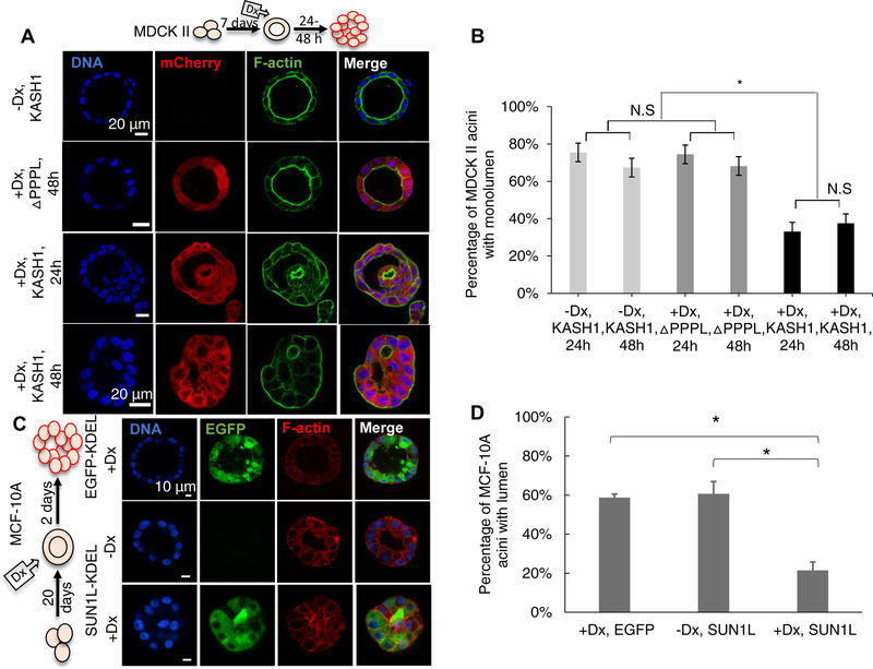
Figure 2. Disruption of the LINC complex induces lumen blockage in pre-formed MCF-10A and MDCK II acini.
Non-induced cells were allowed to form acini; then doxycycline (Dx) was added to induce expression of ΔPPPL: mCherry-KASHΔPPPL and KASH1: mCherry-KASH1 (A) or EGFP-KDEL: SS-EGFP-KDEL and SUN1L-KDEL: SS-EGFP-SUN1L-KDEL (C). The cartoon in (A) and (C) indicates the period of three-dimensional culture (7 d for MDCK II cells, 20 d for MCF-10A cells), and the duration of induction by doxycycline before acinar fixation and microscopy (1–2 d for MDCK II acini, 2 d for MCF-10A acini). (B) Plot shows occurrence of MDCK II acini with single lumen corresponding to the data in (A). n ≥100 acini from 3 independent experiments were scored for each group. Error bars represent ± SEM (*p < 0.001, and N.S. (not significant) by one-way ANOVA followed by Tukey (HSD) test for multiple comparisons). (D) Plot shows the occurrence of MCF-10A acini with lumens under different conditions corresponding to the data in (C). At least 80 acini from 3 independent experiments were scored for each group. Error bars represent ± SEM (*p<0.05 by student’s t-test with Bonferroni corrections for statistical comparisons).

